Ureteric Stone Treatment in Mumbai
A ureteric stone occurs when a urinary calculus drops from the kidney into the narrow ureter, obstructing urine flow and triggering severe, spasmodic flank pain known as acute renal colic.
Modern ureteric stone treatment in Mumbai emphasizes rapid pain relief, precise radiological localization via low-dose CT KUB, and minimally invasive endoscopic resolution. From Medical Expulsive Therapy (MET) for small calculi to incision-free Laser Ureteroscopic Lithotripsy (URSL) and RIRS, patient management is customized to stone size, location, and renal function.
Consult Dr. Shyam Mohan, Consultant Urologist, Uro-Oncologist & Robotic Surgeon in Mumbai, for expert evaluation and evidence-based stone removal.
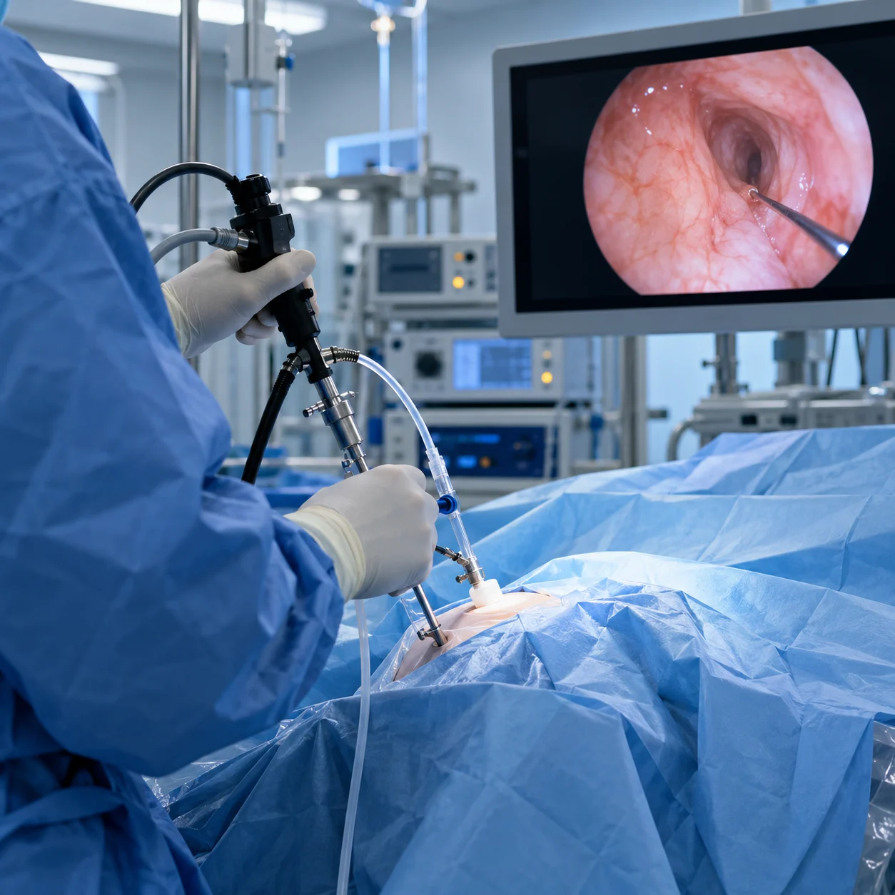
Critical Clinical Alert: Obstructive Pyelonephritis
If ureteric stone pain is accompanied by fever, chills, shivering, uncontrolled nausea, or an inability to pass urine (anuria), immediate emergency hospitalization is required. An obstructed kidney with infected urine can rapidly progress to urosepsis, requiring urgent internal stent decompression.
What is a Ureteric Stone?
The urinary system consists of two kidneys, two ureters, a urinary bladder, and a urethra. The ureters are muscular tubes approximately 25 to 30 cm long and only 3 to 4 mm in internal lumen diameter. Their physiological role is to propel urine from the renal pelvis down into the bladder via involuntary peristaltic contractions.
Virtually all ureteric stones begin as mineral crystal aggregations (composed of calcium oxalate, calcium phosphate, uric acid, or struvite) inside the renal collecting system. When a stone detaches from the kidney and travels downwards, it frequently becomes trapped at one of the physiological narrowing points of the ureter.
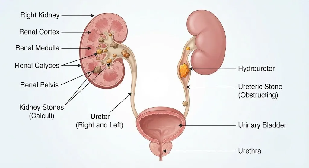 Diagrammatic representation of kidney and
ureteric calculi
Three Common Anatomical Narrowings
Ureteropelvic Junction (UPJ):
Where the renal pelvis transitions into the upper ureter.
Iliac Vessel Crossing:
The mid-ureter segment where the tube crosses over the external iliac blood vessels.
Ureterovesical Junction (UVJ):
The narrowest physiological segment where the ureter enters the muscular wall of the bladder.
When a stone lodges in the ureter, it creates a blockage. Urine continues to be produced above the obstruction, leading to back-pressure, dilation of the ureter (hydroureter), and swelling of the kidney (hydronephrosis). This acute stretching of the renal capsule triggers the excruciating cramping pain characteristic of renal colic.
Symptoms of Ureteric Stones
Unlike silent stones that sit quietly within the renal calyces, a stone moving or stuck in the ureter usually causes acute and unmistakable symptoms:
Severe Renal Colic
Sudden, severe, sharp pain in the flank or lower back that comes in intense waves. Patients frequently cannot find a comfortable sitting or lying position.
Radiating Pain Pathway
As the stone moves lower, pain typically radiates around the abdomen toward the groin, testicles in men, or labia in women.
Hematuria (Blood in Urine)
Urine may appear pink, reddish, or brownish due to the stone's sharp crystalline edges irritating the sensitive ureteral lining.
Urinary Urgency & Frequency
When a stone nears the ureterovesical junction (UVJ) entering the bladder, it triggers intense bladder spasms, frequent urination, and dysuria (burning sensation).
Nausea & Vomiting
Due to the shared celiac autonomic nerve plexus between the kidneys and gastrointestinal tract, renal distension causes persistent nausea and vomiting.
Silent Hydronephrosis
In chronic partial blockages, acute pain can subside into a dull ache while progressive silent kidney damage continues undetected.
Diagnostic Evaluation of Ureteric Stones
Accurate and immediate diagnosis is vital to determine whether a stone can pass naturally or requires active surgical intervention:
| Diagnostic Test | Clinical Purpose | Key Benefit |
|---|---|---|
| NCCT KUB (Non-Contrast CT) | Gold standard imaging. Visualizes the exact stone size, location (upper, mid, or lower ureter), skin-to-stone distance, and Hounsfield density. | Over 98% diagnostic accuracy; detects all stone types. |
| Ultrasound KUB (USG) | Assesses the presence and degree of hydronephrosis (kidney swelling) and identifies stones at the renal pelvis or UV junction. | Radiation-free, safe for pregnant women and initial screening. |
| Urinalysis & Culture | Checks for microhematuria, crystals, leukocyte esterase, and urinary tract infection (UTI). | Identifies concurrent infection that requires urgent management. |
| Serum Creatinine & Electrolytes | Evaluates bilateral renal filtration capacity and assesses whether urinary outflow obstruction has impacted overall kidney function. | Essential for safe surgical planning and medication dosing. |
Treatment Options for Ureteric Stones in Mumbai
Treatment depends on several clinical variables: stone diameter (millimetres), anatomical location, degree of obstruction, severity of symptoms, and whether infection is present.
1. Medical Expulsive Therapy (MET) & Conservative Management
For small stones measuring less than 5 mm located in the distal (lower) third of the ureter without signs of infection or severe hydronephrosis, conservative management is considered safe and effective:
- Selective Alpha-1 Blockers (e.g., Tamsulosin, Silodosin): Relax the smooth muscle fibers in the distal ureter, reducing spasm, widening the lumen, and increasing spontaneous passage rates by up to 30%.
- Targeted Analgesia: Non-steroidal anti-inflammatory drugs (NSAIDs) reduce ureteric inflammation and control colic.
- Adequate Hydration: Controlled fluid intake to maintain urine output without over-distending the obstructed kidney.
- Close Radiological Follow-up: Regular ultrasound review within 2 to 4 weeks to verify stone passage and ensure hydronephrosis has resolved.
2. Ureteroscopic Laser Lithotripsy (URSL / Semi-Rigid URS)
URSL is the globally recognized gold standard intervention for mid and lower ureteric stones, as well as stones larger than 5 to 6 mm that fail conservative passage:
- No Cuts or Scars: An ultra-thin semi-rigid ureteroscope is advanced through the natural urethra and bladder into the ureter under video guidance.
- High-Energy Holmium / Thulium Laser: A hair-thin laser fiber is fired directly against the stone, fragmenting it into fine sand or extractable micro-fragments.
- Grasping Baskets: Dormia nitinol baskets retrieve fragments, clearing the obstruction immediately.
- Rapid Recovery: Day-care procedure performed under short general or spinal anesthesia with minimal postoperative discomfort.
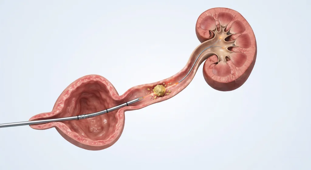 Laser lithotripsy stone dusting
3. Retrograde Intrarenal Surgery (RIRS) for Upper Ureteric Calculi
For stones located in the upper ureter near the kidney, or stones that push back (retropulse) into the renal pelvis during intervention:
A highly flexible, steerable digital ureterorenoscope is maneuvered through the ureter directly into the renal collecting system. The flexible scope can bend up to 270 degrees to reach stones tucked deep inside kidney calyces, fragmenting them completely using advanced laser technology. Learn more on our dedicated RIRS Surgery in Mumbai page.
4. Double-J (DJ) Ureteric Stent Placement
A DJ stent is a thin, flexible tube with curls at both ends—one coiled in the kidney, the other in the bladder:
- Relieves Acute Pressure: Bypasses stone impaction, allowing blocked urine to drain immediately.
- Facilitates Healing: Prevents ureteric swelling and ureteric stricture formation after laser stone fragmentation.
- Quick Outpatient Removal: The stent is temporary and typically removed 1 to 3 weeks later via a 2-minute outpatient cystoscopy procedure.
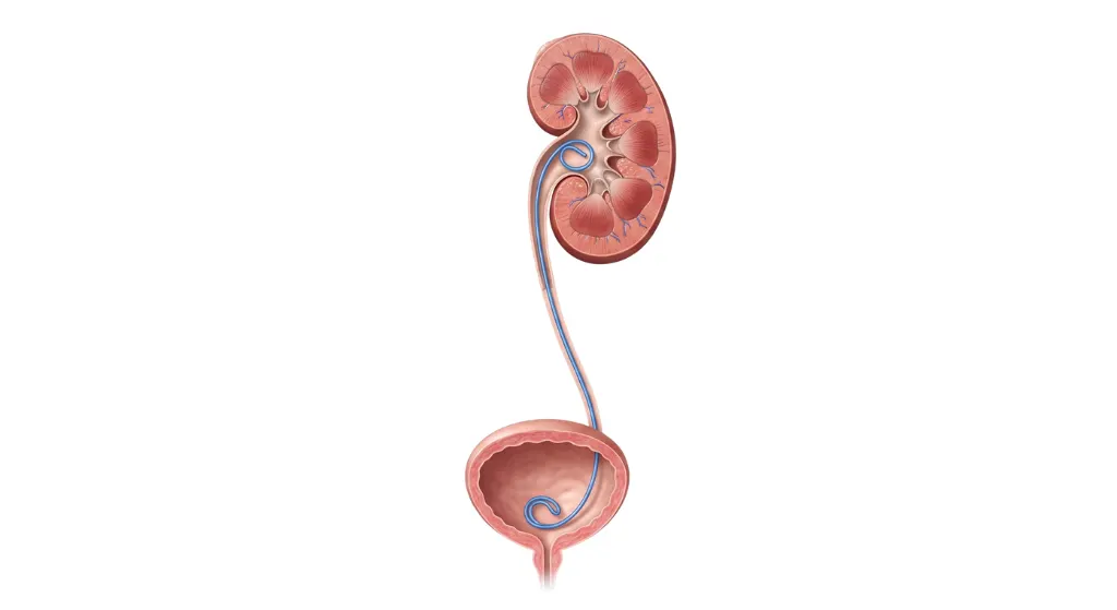 Double-J (DJ) ureteric stent
anatomy
Recovery Timeline After Ureteric Stone Laser Surgery
Day of Surgery
Procedure lasts 30 to 50 minutes. Patients rest in recovery for 2-4 hours, begin oral fluids, and can mobilize gently.
Day 1 to 2
Discharge home. Mild pink urine and mild bladder irritability from the stent are normal and managed with prescribed medications.
Day 3 to 5
Return to office work and routine light activities. Strenuous workouts and heavy weightlifting should be avoided until stent removal.
Week 1 to 3
Outpatient DJ stent removal. Follow-up imaging and 24-hour urine metabolic evaluation to prevent stone recurrence.
Need Expert Evaluation for a Ureteric Stone?
Consultant Urologist, Uro-Oncologist & Robotic Surgeon in Chembur, Mumbai. Complete assessment of acute renal colic, CT scan review, and advanced laser stone removal.
Dr. Shyam Mohan is a highly respected Consultant Urologist, Uro-Oncologist & Robotic Surgeon practicing in Chembur, Mumbai. With extensive fellowship training and clinical mastery in endourology, he routinely performs laser stone surgeries (URSL, RIRS, PCNL) using state-of-the-art Holmium and Thulium fiber lasers, delivering outstanding stone-free rates, minimal postoperative discomfort, and rapid patient recovery.
Urgent & Routine Stone Care: Synergy Health Clinic, Chembur. Same-Day Emergency Consultations Available.
